Bacteria Collections Lab
Part 1
Purpose-to see the growth of bacteria as well as find out how much bacteria are on everyday objects
Hypothesis- That section A which is a computer mouse will have the most bacteria because it is touched by many people many times throughout the day.
Materials-agar plate, Q-tip, objects to swab, inoculating loop, slide, microscope, crystal violet, iodine, ethanol, H20, safrinin, Bunsen burner, test tube, paper circles, bleach, kitchen cleaner, Windex
Method-get an agar plate and q-tips. Divide agar plate into 4 sections. Swab 4 objects and then swab agar plate to transfer bacteria. Place agar plate into incubator to grow bacteria. After a few days, take out bacteria and observe how each section grew and the characteristics of each section. Use inoculating loop to scrape off some bacteria and put on slide. Apply crystal violet and wait for 1-2 mins. rinse with H2O. apply iodine for 1-2 mins. rinse with H2O. wash with Ethanol until purple stops washing away, then rinse with H2O. apply safrinin for 1-2 mins. rinse with H2O and then blot dry. put slide under microscope and observe the colour and forms of bacteria.
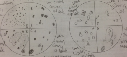
Results-Plate 1, Section A-13 colonies,light yellow and creamy white,translucent, shiny, circular,convex, and entire.
Section B- many colonies, too many to count, creamy white, yellow,translucent, shiny, entire, punctiform, convex
Section C- 15 colonies, yellowish-white, translucent, shiny, entire, circular, and convex
Section D- 30 colonies, yellow, white, grey, translucent, some shiny, some dull, some entire, some lobate, some filamentous, some circular, some irregular, some convex, some flat.
Plate 2-Section E-6 colonies, yellowish-white, translucent, shiny, entire, circular, convex
Section F-12 colonies, yellowish-white, translucent, shiny, entire, circular, convex
Section G-15 colonies, yellowish-white, translucent, shiny,one dull, entire, one undulate, circular, one irregular, convex, one flat
Section H-15 colonies, yellowish-white, one completely white colony, translucent, one opaque, shiny, entire, circular, convex
Analysis-Section D which was the phone in the Biology class was purple after gram staining, so it is gram positive which means it has a thin cell wall with lots of peptidoglycan. It also had three forms of bacteria, coccus, spirilli, and bacilli.
Conclusion-The results did not prove my hypothesis, as section D, which was the phone had the most bacteria growth, and not section A, which was the computer mouse. I might have gotten these results because the phone is also touched by many different people and objects, and probably not cleaned often so bacteria grows well on it. Some further experiments i could do are swabbing more mice in the computer lab and more phones in the classrooms, as then I could have a more accurate comparison as the mouse i swabbed could have been an abnormally clean mouse and the phone could have been an abnormally dirty phone.
Purpose-to see the growth of bacteria as well as find out how much bacteria are on everyday objects
Hypothesis- That section A which is a computer mouse will have the most bacteria because it is touched by many people many times throughout the day.
Materials-agar plate, Q-tip, objects to swab, inoculating loop, slide, microscope, crystal violet, iodine, ethanol, H20, safrinin, Bunsen burner, test tube, paper circles, bleach, kitchen cleaner, Windex
Method-get an agar plate and q-tips. Divide agar plate into 4 sections. Swab 4 objects and then swab agar plate to transfer bacteria. Place agar plate into incubator to grow bacteria. After a few days, take out bacteria and observe how each section grew and the characteristics of each section. Use inoculating loop to scrape off some bacteria and put on slide. Apply crystal violet and wait for 1-2 mins. rinse with H2O. apply iodine for 1-2 mins. rinse with H2O. wash with Ethanol until purple stops washing away, then rinse with H2O. apply safrinin for 1-2 mins. rinse with H2O and then blot dry. put slide under microscope and observe the colour and forms of bacteria.
Results-Plate 1, Section A-13 colonies,light yellow and creamy white,translucent, shiny, circular,convex, and entire.
Section B- many colonies, too many to count, creamy white, yellow,translucent, shiny, entire, punctiform, convex
Section C- 15 colonies, yellowish-white, translucent, shiny, entire, circular, and convex
Section D- 30 colonies, yellow, white, grey, translucent, some shiny, some dull, some entire, some lobate, some filamentous, some circular, some irregular, some convex, some flat.
Plate 2-Section E-6 colonies, yellowish-white, translucent, shiny, entire, circular, convex
Section F-12 colonies, yellowish-white, translucent, shiny, entire, circular, convex
Section G-15 colonies, yellowish-white, translucent, shiny,one dull, entire, one undulate, circular, one irregular, convex, one flat
Section H-15 colonies, yellowish-white, one completely white colony, translucent, one opaque, shiny, entire, circular, convex
Analysis-Section D which was the phone in the Biology class was purple after gram staining, so it is gram positive which means it has a thin cell wall with lots of peptidoglycan. It also had three forms of bacteria, coccus, spirilli, and bacilli.
Conclusion-The results did not prove my hypothesis, as section D, which was the phone had the most bacteria growth, and not section A, which was the computer mouse. I might have gotten these results because the phone is also touched by many different people and objects, and probably not cleaned often so bacteria grows well on it. Some further experiments i could do are swabbing more mice in the computer lab and more phones in the classrooms, as then I could have a more accurate comparison as the mouse i swabbed could have been an abnormally clean mouse and the phone could have been an abnormally dirty phone.
